Sunday, June 5, 2016

ADOPTED MICROBE !

Hi and Greetings !

          On 3rd June 2016, I’ve presented my adopted microbe which is Cupriavidus metallidurans. Of course, before the presenting, I have to search and prepared a lot of things including reading, searching and understand it clearly and finally do a scrapbook about it.

          I’ve chosen this microbe because it was really impressed me where C.metallidurans eats toxis materials and then.... well.... POOPS GOLD ! Awesome right ?! It is also a Gram-negative , motile, non-spore forming, rod-shaped bacterium known for its ability to resist toxic heavy metals; metallidurans literally translates to "metal-enduring". C. metallidurans is normally found in industrial sediments or wastes which contain high heavy metal concentrations. This microbial magician named Cupriavidus metallidurans , when placed in a minilab full of gold-chloride (a nasty toxin) gumble up the poison and in about a week process it out as 24-karat of precious yellow metal which is GOLD.
The bacteria was found to be 25 times more resistance to the metal based toxin chemical. Metal resistance functions are predominantly encoded on two plasmids, pMOL28 and pMOL30, which produce metal exporters that pump metal ions out of the cell, thus protecting intracellular macromolecules from the toxic effects of high concentrations of metal. It is also believed helping our environmental cause and making us a small profit in the process.

          I’ve searched anywhere in the internet and at the same time finished my scrapbook based on above content :
  • Scientific name
  • Images
  • Classifications
  • Description
  • Ecology
  • Metabolism and Nutrition
  • Significant
  • References

          This is assignment on  adopting microbes is to present a 3 minutes thesis as well as to learn more and understand the microbes is. I also have been working on a digital scrapbook  which I prefer on using PowToon.

Want to know more about my microbe ?? Click here >> "Cupriavidus metallidurans"


This was the picture of my 3MT presentation.

3MT presentation picture.


From the 3 minutes presentation, I’ve also learnt and gained my confident to talk infront of many peoples eventhough I’m a little bit of shaking. HAHA!

In short, I did really enjoy on the this assignment because it add-on my experiences and also attracted me to want to learnt it more.

YAKULT’S FACTORY TRIP

Hi and Greetings !

Malaysia's Yakult Factory


On 16 May 2016, We had a trip to Yakult’s Factory. The factory was located at Seremban and it takes one hour to reached the destination. We was departed from UPM at 8.30am and arrived at 9.30am by bus. As we arrived safely, we’ve been asked to directly entered the Main Hall to attend some presentation or briefing on what is the factory do.
 Malaysia’s Yakult Factory

Lactobasillus Casei Shirota
 It is a factory that made cultured drink by using a bacteria that is very useful in food production such as YAKULT that gives benefits to the worldwide. The factory also  involves in food production and packaging. YAKULT is a high quality probiotics in the form of a cultured drink. It contains the probiotic bacteria "Lactobacillus casei Shirota" and also know as the "Shirota strain". With over 30 billion live Shirota strain in each bottle, YAKULT has among the highest concentrations of probiotics compared to other probiotic products on the market. The Shirota strain is scientifically proven to be the strongest strains af beneficial bacteria, and has been shown to benefit human health. It also free of preservatives, colourings and stabilizers, so YAKULT is the right choice for those looking for high quality of probiotics for good health. There are two type of YAKULT; Yakult Ace and Yakult Ace Light.


Left : Yakult Ace, Right : Yakult Ace Light

Yakult Ace used sucrose while Yakult Ace Light used fructose which is less sugar than Yakult ace.


After we had been given the talk, we were brought to  see the process of packaging of YAKULT step by step. There were many phases, stages and processes involved in packaging the YAKULT's bottles and they make sure that there was no defects in the packaging. 

Process of making YAKULT.
Packaging process.


Some of the pictures :


Some of the pruduct that used Shirota strain.
While on the road to Yakult's Factory
Candid :)
All of us including Dr. Fairol.


CHAPTER 9 - PRINCIPLES OF MICROBIAL ECOLOGY

Hi and Greetings !

          For this class the learning concept as a bit differ than before whereas we need to teach our friend about this topic. This activity occurs in the group where the every person in the group have decided on what he or she wants to explained to the other group members. Until the last group, they will gained an extra information about the this topic. At the same time, that last group need to explain it again to the first group where the first group will gained another extra information about this topic.
So, when my group turn up to give an explanation to the next group, because of my group members only 3 person attended the class that time, so each of us got 2 subtopics to be explained to the next group.  

          I got the subtopics about Levels of Organization and also Homeostasis. This activity has taught me on how to deliver the information clearly and effectively to other person and it really gave me a satisfaction when they got the info clearly based on the subtopics. The moment I give an explanations, I got not enough of time to explain because I need to explain to two person at that time, so I need explain one subtopic by one subtopic.

          Overall, this week class really gave us so many experiences on explaining something in facing each others. At the end of the class, the lecturer ask on what subtopic that we not clear and then the lecturer explained it clearly to us.


          Last but not least, we’ve been asked to make a summary based on this chapter :


Sunday, April 17, 2016

VISITING IBS UPM (INSTITUTE OF BIOSCIENCE)

Hi and Greetings !

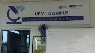
On Friday 25th March 2016, we got an opportunity to visited IBS (Institute of Bioscience) to study, identify and also carry out on how they conducting an experiment under different types of microscope. There were various types of microscope used including :
  • Transmission Electron Microscope (TEM)
    • works like slide project : light transmit through slide, light passes through slide, light passes through as affected by structure and objects on slides (different intensity passes through), image enlarge on a viewing screen.
    • usually used to observe internal structure of cell that is very thin layer and cross section.
  • Scanning Electron Microscope (SEM)
    • highly similar to TEM sample except sectioning
    • fixed, dehydrated, mounting and coating
    • usually used to observe surface structure of cell

Figure below shows some of the picture I took in our visit to IBS :


Overall, 

CHAPTER 4 - EUKARYOTIC CELLS

Hi and Greeting viewers !


So this is all about topic 4 of the lecture that is Eukaryotic Cells. This topic was actually almost the same with the previous lecture that about Prokaryotic Cells but some of the organelles or structures functions were slightly different. But, some of the organelles or structure of eukaryotic cells were absent in prokaryotic cells.


Figure 1 : Similarities and Differences between Prokaryotes and Eukaryotes


As we can see on Figure 1 that there was a similarities and differences between prokaryotes and eukaryotes. Not only that, it was also have different functions every each of the structures but not all off it because there was also the structure that have the same function as prokaryotes.

On this lecture, Dr. Wan gave us a group presentation which is in the kind of games such as puzzle, flip-a-card and many more. We had been assigned in 6 groups where each of the group have to conducted a different types of games. I was in the Group 4 where we have decided to make Flip-a-card games. Our games came out with rules to make it happening and not boring. But there was a group that gave a punishment to any groups who answered it wrong. It was really happy for us and I am the one who volunteer to accompany the groups that got the punishment. One of the punishment is :


Link : One of the punishments to a groups who answered a question wrongly.


As a conclusions, based on the games conducted about Eukaryotic cells, I have gained many output including able to list down and identified the important structure of Eukaryotic cells, able to explained the characteristics or functions of the structures of Eukaryotic cells and also able to describe the process of meiosis and mitosis.

CHAPTER 3 - PROKARYOTIC CELLS

Hi and Greetings !

So, this time I’ll talk about topic 3 that is Prokaryotic cells. In this 3 times lectures, I have gained many information and knowledge about prokaryote. In fact, I have gained so much experience on the activities conducted by Dr. Wan herself. It was very awesome !
It all starts from the word “microorganism”. We have known that Microbiology is the study of small living things. But is there life in microorganisms? So, to identify it, there were biologist state that life must have at least the following processes, that was :
  • Growth
  • Reproduction
  • Responsiveness
  • Metabolism
  • Cellular Structure


Figure 1 : Similarities and Differences between Prokaryotes and Eukaryotes


So what is Prokaryotic Cell?
  • It is include bacteria and Achaea
  • Size range from 0.2 – 2.0 um (diameter) and 2 – 8 2 um (length)
  • Lack of :

o   Membrane-bound nucleus
o   Cytoskeleton
o   Membrane-bound organelles
o   Internal membranous structures


There were also many shapes of bacteria I have ever studied as shown on the Figure 2 below.


Figure 2 : Bacterial Shapes and Arrangements


I also have identified on what makes a species to have a particular size and shape. This was caused by the efficient of nutrient intake and also the decreased susceptibility of predation.

Other that, after learnt about prokaryotic cells, I able to identify the important structures and also able to explain the characteristics of functions of the structures of prokaryotic cells.


Link : Summary of important structures and some explanation of the characteristics of function of prokaryotic cells


Based on the link given, that was one of the activities that Dr. Wan asked us to do. At the beginning of this topic, she asked all of us to draw a mind map about prokaryotes on the whiteboard because she already told us to study this topic first before attending the class. And the result as we can see from the Figure 4.


Figure 4 : Mind map drew by us.


Other that, I have also learnt about some of the inclusion and it’s type, including :
  • Polysaccarides granules
  • Lipid inclusions
  • Sulfur granules
  • Carboxysome
  •  Gas vacuoles
  • Magnetosomes


Overall, I have gained so much information much better about what is prokaryotic cells including able to identified the  important structures of prokaryotic cells and also able to explain the characteristics of functions of the structures of prokaryotic cells. 

CHAPTER 2 - MICROSCOPY

Hi and Greetings viewers !
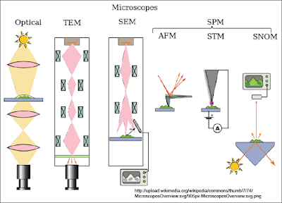
This is topic 2 for this subject that is Microscopy. On what have I learnt in this topic, there were 4 types of microscope including :
  • Optical Microscope, that includes :
    • Dark field
    • Bright field
    • Fluorecence microscopy and
    • Phase-contrast microscopy
  • Electron Microscope, that includes :
    • Scanning Electron Microscope (SEM)
    • Transmission Electron Microscope (TEM)
    • Environmental Scanning Electron Microscope (ESEM)
  • Scanning Probe Microscope, that includes :
    • Atomic Force
    • Scanning Tunnelling
    • Scanning Near-Field Optical Microscope, and
    • Etc
  • Scanning Acoustic  Microscope


Figure1 above shows some types of the microscope characteristics.

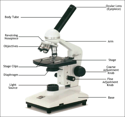
In this topic I have also learnt on how to identify the parts of basic microscope as we can see on the Figure 2.


                                                     Figure 2 : Basic Microscope Parts


Gram positive bacteria are “positively” stained purple/blue when chemicals of the gram staining process bind to them. Gram negative bacteria on the other hand respond “negatively” to the gram staining treatment. So they have a barrier preventing the gram staining procedure from turning it blueish purple.


Figure 3 : Gram Positive and Gram Negative


I also can recognize images produced from different microscope and how to select the suitable microscope for relevant usage. For an examples :


Figure 3 : Bright-Field Microscope


Figure 4 : Dark-Field Microscope


Figure 5 : Phase-Contrast Microscopy


Figure 6 : Fluorecence Microscopy


Figure 7 : Transmission Electron Microscope (TEM)


Figure 8 : Scanning Electron Microscope (SEM)


I can also describe the principle of light, phase-contrast, fluorescence and electron microscope, where :
  • Bright-Field Microscope is for enhanced observation with bright-field microscope,kill and stain the cells
  • Dark-Field Microscope is usually used to observe live specimens which cannot be stained. Only light that is reflected off the specimen enters the objective lens.
  • Phase-Contrast Microscope permits detailed examination of internal structures of the cells.
  • Normaski Differential Interference Contrast (DIC) Microscopy creates a 3D image of the specimen.
  • Fluorecence Microscope produced image from light that passes through a specimen where it naturally fluorescing against dark background.
  • Confocal Microscpy used for imaging thick specimens with many planes of reflection.
  • Transmission Electron Microscope basically used for analysing internal structures of the cells.
  • Scanning Electron Microscope basically used for analysing surface structures of the cells.
  • Atomic Force Microscopy allows observation and manipulation at molecular and atomic level.
  • Scanning Atomic Microscopy usually used to studying larger specimens like bacterial biofilms, cancel cells and others.


Dr. Wan Zuhainis also taught us this topic by doing some activity such as short-quizzes on indentifying the types of microscope by the picture shown on the slides.