Hi and Greetings viewers !
This is topic 2 for this subject that is Microscopy.
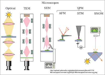
On what have I learnt in this topic, there were 4 types of microscope including
:
- Optical Microscope, that includes :
- Dark field
- Bright field
- Fluorecence microscopy and
- Phase-contrast microscopy
- Electron Microscope, that includes :
- Scanning Electron Microscope (SEM)
- Transmission Electron Microscope (TEM)
- Environmental Scanning Electron Microscope (ESEM)
- Scanning Probe Microscope, that includes :
- Atomic Force
- Scanning Tunnelling
- Scanning Near-Field Optical Microscope, and
- Etc
- Scanning Acoustic Microscope
Figure1 above shows some types of the microscope characteristics.
In this topic I have also learnt on how to identify
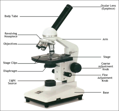
the parts of basic microscope as we can see on the Figure 2.
Figure 2 : Basic Microscope Parts
Gram positive bacteria are “positively” stained
purple/blue when chemicals of the gram staining process bind to them. Gram
negative bacteria on the other hand respond “negatively” to the gram staining
treatment. So they have a barrier preventing the gram staining procedure from
turning it blueish purple.
Figure
3 : Gram Positive and Gram Negative
I also can recognize images produced from different
microscope and how to select the suitable microscope for relevant usage. For an
examples :
Figure
3 : Bright-Field Microscope
Figure
4 : Dark-Field Microscope
Figure
5 : Phase-Contrast Microscopy
Figure 6 : Fluorecence Microscopy
Figure
7 : Transmission Electron Microscope (TEM)
Figure
8 : Scanning Electron Microscope (SEM)
I can also describe the principle of light,
phase-contrast, fluorescence and electron microscope, where :
- Bright-Field Microscope is for enhanced observation with bright-field microscope,kill and stain the cells
- Dark-Field Microscope is usually used to observe live specimens which cannot be stained. Only light that is reflected off the specimen enters the objective lens.
- Phase-Contrast Microscope permits detailed examination of internal structures of the cells.
- Normaski Differential Interference Contrast (DIC) Microscopy creates a 3D image of the specimen.
- Fluorecence Microscope produced image from light that passes through a specimen where it naturally fluorescing against dark background.
- Confocal Microscpy used for imaging thick specimens with many planes of reflection.
- Transmission Electron Microscope basically used for analysing internal structures of the cells.
- Scanning Electron Microscope basically used for analysing surface structures of the cells.
- Atomic Force Microscopy allows observation and manipulation at molecular and atomic level.
- Scanning Atomic Microscopy usually used to studying larger specimens like bacterial biofilms, cancel cells and others.
Dr. Wan Zuhainis also taught us this topic by doing
some activity such as short-quizzes on indentifying the types of microscope by
the picture shown on the slides.


No comments:
Post a Comment